The Multiple Myeloma Channel is supported with funding from Sanofi (Gold) and Legend Biotech (Bronze).
VJHemOnc is an independent medical education platform. Supporters, including channel supporters, have no influence over the production of content. The levels of sponsorship listed are reflective of the amount of funding given to support the channel.
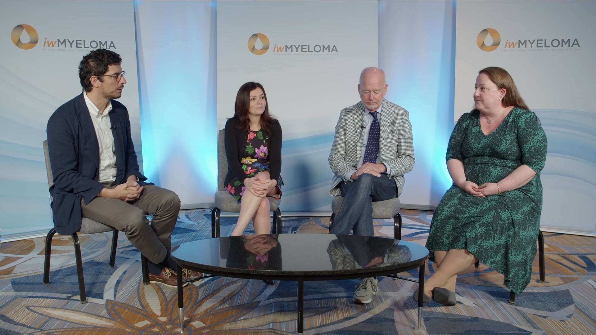
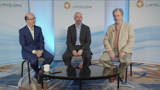
iwMyeloma 2026 | Advances in myeloma genomics: characterizing structural variation and identifying prognostic factors
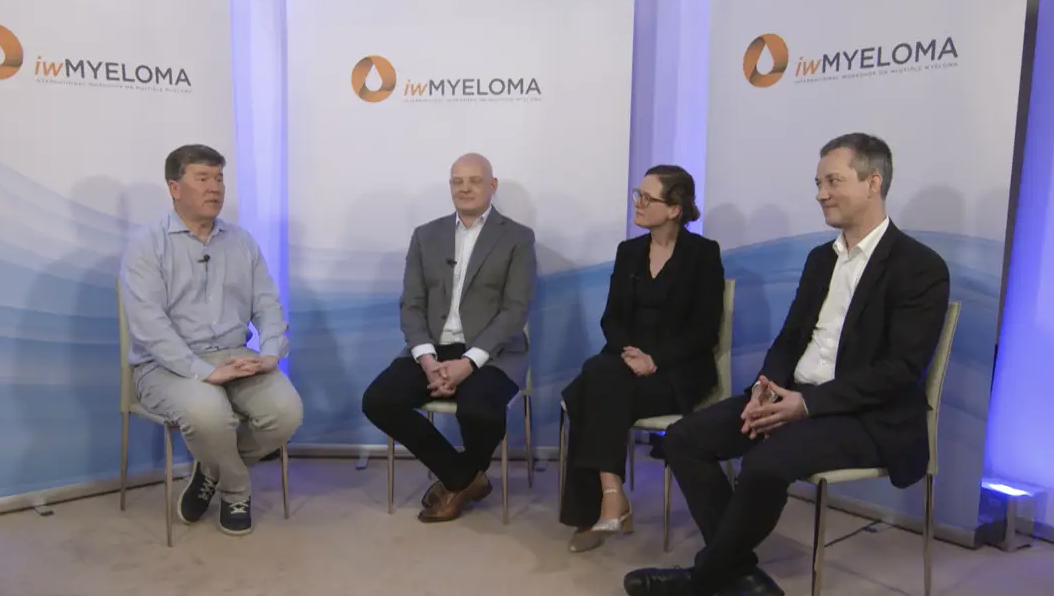
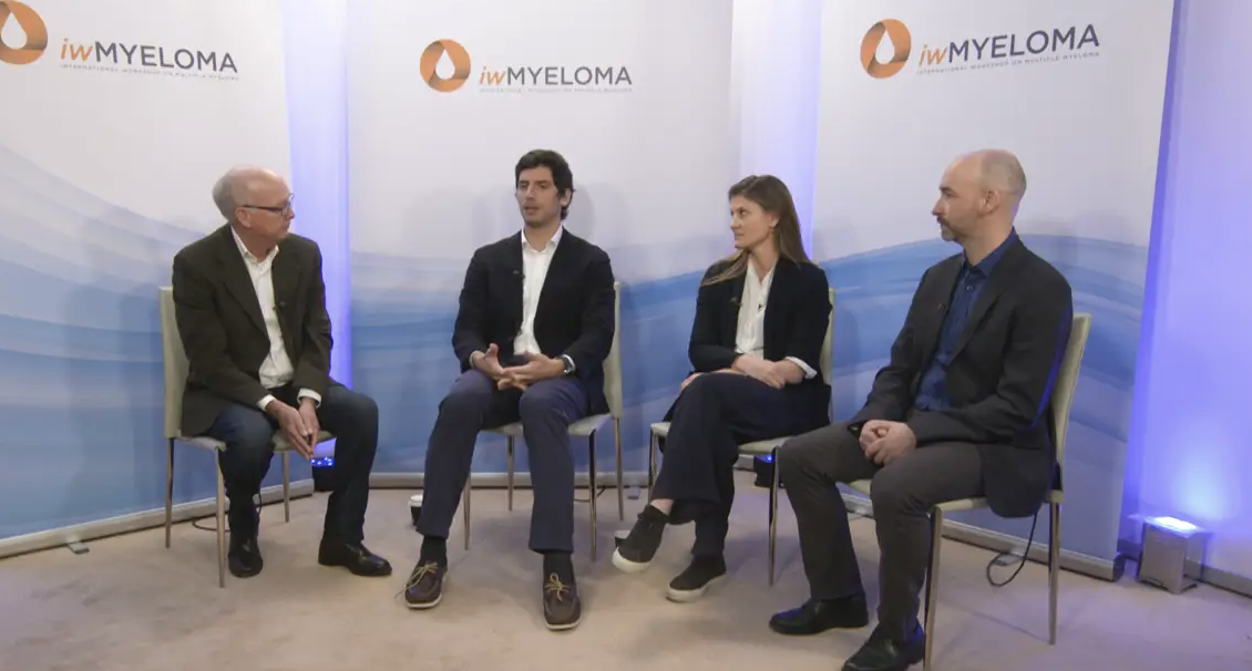
In this discussion, Leif Bergsagel, MD, Mayo Clinic, Phoenix, AZ, Niels Weinhold, PhD, University Hospital Heidelberg, Heidelberg, Germany, Mehmet Samur, PhD, Dana-Farber Cancer Institute, Boston, MA, Brian Walker, PhD, Sylvester Comprehensive Cancer Center, University of Miami, Miami, FL, and Patrick Blaney, MS, Perlmutter Cancer Center, NYU Langone Health, New York, NY share updates on the genomics of multiple myeloma (MM). The experts discuss the value of computational methods and multi-omic sequencing approaches for characterizing and interpreting genetic alterations and potentially improving risk stratification in the future. Dr Weinhold provides insight into the biology of bone marrow-independent disease and highlights the immune heterogeneity across myeloma lesions, which limits the ability to reliably predict treatment response. This discussion was filmed at the 19th International Workshop on Multiple Myeloma (iwMyeloma), held in Miami, FL.
These works are owned by Magdalen Medical Publishing (MMP) and are protected by copyright laws and treaties around the world. All rights are reserved.
Transcript
Leif Bergsagel:
Hi, I’m Leif Bergsagel from the Mayo Clinic in Arizona, and I’m here at iwMyeloma 2026. We just finished a really exciting session on the genomics of multiple myeloma, and I’m joined by my panel here.
Niels Weinhold:
Niels Weinhold from Heidelberg...
Leif Bergsagel:
Hi, I’m Leif Bergsagel from the Mayo Clinic in Arizona, and I’m here at iwMyeloma 2026. We just finished a really exciting session on the genomics of multiple myeloma, and I’m joined by my panel here.
Niels Weinhold:
Niels Weinhold from Heidelberg.
Mehmet Samur:
Mehmet Samur from Dana-Farber.
Brian Walker:
Brian Walker from University of Miami.
Patrick Blaney:
Patrick Blaney from NYU Langone Health.
Leif Bergsagel:
Okay. So maybe Patrick, we’ll start with you. You know, you presented a really important topic, which is we have all these genomic changes in multiple myeloma, particularly structural variations, but they’re so complicated that it’s very hard to compare what’s been done in different studies or even between different patients. But you’ve come up with a way that’s going to help simplify that. Do you want to expand on that?
Patrick Blaney:
Yeah, so genome graphs are a kind of computational approach that allows us to integrate both copy number and structural variation, which up until recently has kind of been done via manual annotation, which that’s a wonderful approach, but it tends to be difficult to reproduce. It requires a lot of training and a lot of time dedicated by individuals to be able to piece these things together. So finding an alternative that integrates things that we’ve already generated so copy number data and structural variation but then applying an additional computational method that does a lot of the legwork and is able to produce and reconstruct these very complex genomes is I think essential in trying to move towards a more consistent basis of defining structural variation and in particular defining complex events. And so, I think that it’s exciting for us to be able to kind of get this data set fully analyzed, but also to see what kind of interesting kind of derivative chromosomes are available for us to investigate and maybe contribute to high risk more than other derivative chromosomes. Not all complex events are created equal, but some of them are likely to be more recurrent than others, I believe.
Leif Bergsagel:
And do you see differences in that score between the two main types of myeloma, hyperdiploid and myeloma with translocations?
Patrick Blaney:
Yeah, so I think that preliminarily, I actually think that the hyperdiploid group might have more chromothripsis and more complex events than others. Now, is that because there’s more chromosomal material available? That’s a good question. I, you know, maybe we can, I can answer that later on with some more analysis, but yeah, I do believe that there is going to be a difference between them, but, trying to see if there are particular sites that contribute much greater to the phenotype is going to be a bit of a difficult process. But I think that we have the data to be able to do it and then to do it at an individual disease stage approach and not necessarily have to do it across the entire spectrum of myeloma.
Brian Walker:
So kind of like interestingly, so we kind of looked at the CoMMpass data set for chromothripsis events. We saw that there was an enrichment actually in the t(4;14) patients who have del(17p). So it seems to be again kind of linked to those high-risk groups in some way as well.
Leif Bergsagel:
Actually, it brings us to your presentation. The micro-C, I mean, you did multi-omic analysis that looked at structural variations at the genetic marks, the micro-C. I thought that was really interesting the way you could delineate the chromothripsis events and significance. Do you want to expand a bit on that?
Brian Walker:
Yeah, so we’ve been using our patient-derived xenograft models to do multi-omics. So most of the kind of patient data out there is standard Illumina whole genome sequencing and RNA sequencing, but we’ve been doing newer third-generation sequencing, which gives us longer reads, as well as the micro-C, as you said, and then also looking at histone marks as well so that we can look at the epigenetics of these samples and being able to kind of add in the micro-C and the epigenetic marks allows us to see how those chromothripsis events are being rearranged and then also how sometimes they get duplicated onto secondary chromosomes as well, and can affect those by causing gains and deletions there. We also kind of see that the epigenetics of these chromothripsis regions changes as well, and that kind of process ends up in dysregulating key genes such as MYC through juxtaposition of those super enhancers as well, yet another mechanism causing gene dysregulation of some of our known targets as well.
Leif Bergsagel:
I guess it’s fair to say that these aren’t advances that are likely to be moved forward into the clinic, but they’re really things that are going to help us in the basic biology to understand. Or do you think any of this would ever move forward to the clinic?
Brian Walker:
It’s an interesting question. So, I think with some of the micro-C, they’re getting down to small numbers of cells that you can do this. And interestingly, you can get structural variants, mutations, and those interactions all from that micro-C data. And so, it could be an alternative to standard whole genome sequencing. Again, the limitation is moving any of these sequencing technologies into the clinic, which is quite difficult as it is anyway.
Leif Bergsagel:
Mehmet, what you presented with the role of the different genetic events and progression clearly has got a lot of clinical importance. Do you want to expand on your presentation?
Mehmet Samur:
Yeah, I mean, as we are looking at these precursor conditions, obviously we see genomics play a role on risk stratification even early on. And in some aspects, they are very similar to myeloma. Between the progressors and non-progressors, we see clear differences as well. So, it adds to the point that we can use this information clinically to stratify the patients. But as Patrick and Brian was also discussing, there are additional information other than DNA that we can add on top of it. Brian was showing, for example, the epigenetic regions or the transcriptomic changes. We have been seeing data coming out on the proliferation and you and I was discussing, like, do we need to do RNA seq to look at the proliferation? Maybe not. We can come with much easier assays to look at it. But as we combine all this different information with one assay or another, I think there is definitely a clinical utility to it. What also makes me very excited from Brian’s and Patrick work is that we used to take everything as a linear because we take the genome as a linear piece, and then we see two events too far apart from each other. But data clearly shows that we should get away from this linear approach, and then everything actually may be much closer than what we think on the three-dimensional space, and then they may be linked to each other. I think as we accumulate more and more, we will learn definitely more and I’m certain that this will eventually lead us to some meaningful clinical transformation at certain point.
Leif Bergsagel:
And Niels, you know, whereas Patrick and the others have sort of looked at a tumour from many different angles, you’ve looked at multiple tumours and their microenvironment to make everything more complicated, but showing that the disease and the microenvironment is so different in the different areas. You want to expand on that?
Niels Weinhold:
Yeah. So, in my presentation, at least, I tried to show that based on our studies in the last decade, there might be targets for treatment, so that specifically target the so-called bone marrow independent loci. So this includes focal lesions that just left the bone marrow, so these so-called breakout lesions, but at the same time, these are also the extramedullary lesions. And our studies of genomics of these lesions have shown that they’re usually highly advanced, but we haven’t identified any kind of uniform driver yet. So that makes it a bit complicated to directly target the tumor cells. However, what we observed and what we have described for the first time in myeloma is that even in newly diagnosed patients, we see signatures of exhaustion in T-cells that have also been described in the solid tumor field. And there might be a therapeutic option here, but at the same time, I showed that there is extensive intra- and interlesional heterogeneity. So there are lesions with hardly any T-cells. There are other lesions that are full of T-cells, but don’t show any signatures of exhaustion. And we do see these lesions with signatures of exhaustion. We also investigate that in the context of immunotherapies and show that two lesions, both of them with signatures of exhaustion, one responds the other one does not respond so that means in the end it’s not that easy just based on single parameters to predict response to treatment and also define these signatures as targets for new treatments so, in principle as you said it’s it a complex story. It makes it even more complex. So, the conclusion is, so either we spend even more work into a better understanding of the biology of these lesions, or I also mentioned an alternative, we can also try to prevent the development of these lesions so we don’t even have to deal with these features.
Leif Bergsagel:
So, if I’m a patient and my doctor tells me I’ve got exhausted T-cells, what should I do?
Niels Weinhold:
Thank you, Leif. So, yeah, so there are these so-called checkpoint inhibitors that can target these T-cells, but usually they don’t target these terminally exhaustive T-cells, but precursors of these T-cells. And we also do see them in these myeloma lesions. But as you know, so the first attempts to use these inhibitors, I guess it’s more than 10 years ago in myeloma. So it was a total failure. So no clinical benefit at all. However, at that time, I guess there was no distinction between bone marrow disease and bone marrow independent disease. So it could be that there was some kind of benefit, but actually we don’t know. And as I said, so we see these examples, exhausted T cells only, or at least showing signatures of exhaustion. And after one week of treatment with a bispecific antibody, the lesion is gone. So just because of the presence of these T cells, it does not mean that the therapy will not work.
Mehmet Samur:
From the translational point, do you think those T-cells that we detect around the tumor cells can also help us to figure out which T-cell phenotype may be more useful to generate CAR T-cells? You know, we discuss a lot about CD4 positive cells, CD8 positive cells, by which phenotype to follow. If they’re more fighting with the tumor cells, do you think they won’t be a better candidate to at least study?
Neils Weinhold:
So I don’t have much experience in that field. But however, looking from the opposite side, so we do see the signatures of exhaustion within lesions. And at the same time, the signatures of exhaustion also suggest that these T-cells are directed against these tumor cells. So that means the data we have already collected could help us to identify the targets of these T-cells and based on these findings, then define new targets for treatment. But it’s also a long wait to go. But as I said, so these T-cells are clearly directed against tumor cells. So the next step will be to get a better understanding. So first, what triggers these immune responses within lesions and what are the targets of these T-cells? And then we could have also some clinical benefit.
Leif Bergsagel:
Well, that’s great. Well, there you have it. A very exciting session to open up the iwMyeloma 2026 in Miami.
This transcript is AI-generated. While we strive for accuracy, please verify this copy with the video.
Read more...